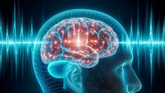
The brain's natural waste-clearing process becomes less effective during sleep as people age, according to a new MIT study. Researchers found that older adults have weaker waves of cerebrospinal fluid, linked to changes in brain activity and blood flow, potentially offering new avenues for research into cognitive decline and dementia.
Researchers at MIT have found that waves of cerebrospinal fluid, which help clear waste from the brain during sleep, become weaker with age.
As people age, they may sleep for shorter periods and wake up more frequently during the night. In addition to those disruptions, MIT researchers have now discovered that a process responsible for flushing waste from the brain during sleep is commonly impaired in older people.
The researchers found that waves of cerebrospinal fluid (CSF), which help remove debris from the brain, are significantly reduced in older adults. This impairment is linked to a decrease in the slow brain waves that normally occur during deep sleep, the researchers found, and is not just a result of getting less sleep.
The researchers now hope to explore whether the disruption of CSF flow increases the risk of neurodegenerative disorders, and if any of the sleep patterns they observed might offer a way to predict an individual’s risk.
“We know that people age really differently. Some people age very successfully with great cognitive outcomes, and other people develop cognitive decline. If we can figure out who has great trajectories of aging and what are the factors that are associated with that, we might be able to use that to predict future outcomes,” says Laura Lewis, the Athinoula A. Martinos Associate Professor of Electrical Engineering and Computer Science, a member of MIT’s Institute for Medical Engineering and Science and the Research Laboratory of Electronics, and an associate member of the Picower Institute for Learning and Memory.
Her lab also plans to investigate whether there might be any interventions that could improve CSF flow in older adults.
Lewis is the senior author of the study, which appears today in Nature Aging. Sydney Bailes, a postdoc in the MIT Laboratory for Information and Decision Systems, and Stephanie Williams, a former visiting student at MIT who earned her PhD from Boston University in 2026, are the lead authors of the paper.
Smaller Waves
During the deepest stages of sleep, also called non-REM sleep, high-amplitude, low-frequency waves contribute to physical recovery, memory consolidation, and many other functions. In a 2019 study, Lewis found that these slow waves are also critical for generating waves of CSF that help flush waste from the brain.
That finding led her to wonder whether reductions in slow waves in older people might also reduce CSF flow. Previous studies of aging have yielded conflicting evidence; some found that CSF flow goes down in older adults, while others found it goes up, but these studies did not monitor flow during sleep.
In the latest study, the researchers recruited volunteers in two age groups: 18 to 39 and 60 to 84. The tests lasted for about two hours and began late at night, so that participants would be sleepy.
The researchers used traditional fMRI to measure participants’ blood flow in the brain, along with a modified MRI technique, developed by Lewis, that measures CSF flow. While the subjects were in the scanner, the researchers also measured their brain waves using EEG.
Many of the participants fell asleep in the scanner, allowing the researchers to compare their brain activity while awake and while sleeping.
As they had seen in earlier work, the researchers found that when the younger adults fell into non-REM sleep, their EEG slow waves became stronger, and their CSF waves became larger. However, in the older group, the average CSF waves were significantly smaller than those seen in the younger group.
Impaired Flow
In additional experiments done while subjects were fully awake, the researchers tried to figure out what was causing the age-related declines in CSF flow.
First, they explored the possibility that changes in a phenomenon known as neurovascular coupling might play a role. When localized brain activity increases the demand for oxygen, nearby blood vessels dilate and deliver more oxygen and nutrients. That increase in blood flow can be measured with fMRI.
Date: 08.12.2025
Naturally, we always handle your personal data responsibly. Any personal data we receive from you is processed in accordance with applicable data protection legislation. For detailed information please see our privacy policy.
Consent to the use of data for promotional purposes
I hereby consent to Vogel Communications Group GmbH & Co. KG, Max-Planck-Str. 7-9, 97082 Würzburg including any affiliated companies according to §§ 15 et seq. AktG (hereafter: Vogel Communications Group) using my e-mail address to send editorial newsletters. A list of all affiliated companies can be found here
Newsletter content may include all products and services of any companies mentioned above, including for example specialist journals and books, events and fairs as well as event-related products and services, print and digital media offers and services such as additional (editorial) newsletters, raffles, lead campaigns, market research both online and offline, specialist webportals and e-learning offers. In case my personal telephone number has also been collected, it may be used for offers of aforementioned products, for services of the companies mentioned above, and market research purposes.
Additionally, my consent also includes the processing of my email address and telephone number for data matching for marketing purposes with select advertising partners such as LinkedIn, Google, and Meta. For this, Vogel Communications Group may transmit said data in hashed form to the advertising partners who then use said data to determine whether I am also a member of the mentioned advertising partner portals. Vogel Communications Group uses this feature for the purposes of re-targeting (up-selling, cross-selling, and customer loyalty), generating so-called look-alike audiences for acquisition of new customers, and as basis for exclusion for on-going advertising campaigns. Further information can be found in section “data matching for marketing purposes”.
In case I access protected data on Internet portals of Vogel Communications Group including any affiliated companies according to §§ 15 et seq. AktG, I need to provide further data in order to register for the access to such content. In return for this free access to editorial content, my data may be used in accordance with this consent for the purposes stated here. This does not apply to data matching for marketing purposes.
Right of revocation
I understand that I can revoke my consent at will. My revocation does not change the lawfulness of data processing that was conducted based on my consent leading up to my revocation. One option to declare my revocation is to use the contact form found at https://contact.vogel.de. In case I no longer wish to receive certain newsletters, I have subscribed to, I can also click on the unsubscribe link included at the end of a newsletter. Further information regarding my right of revocation and the implementation of it as well as the consequences of my revocation can be found in the data protection declaration, section editorial newsletter.
The researchers tested each subject’s neurovascular coupling by measuring blood flow while the subjects looked at a flickering checkerboard image — a task that typically drives an increase in neural activity, along with an increase in blood volume.
In many of the older subjects, while their neural activity did go up, the blood flow didn’t increase much, suggesting that the blood vessels’ ability to respond to increased oxygen demand has diminished.
During another task, the researchers tested whether the brain’s vascular system might be impaired in the older subjects. They asked the subjects to hold their breath, then measured what happened inside the brain. Normally, this leads to an increase in carbon dioxide levels in the blood, which triggers an increase in cerebral blood flow. However, in the older participants, the average blood flow increase was smaller than the increase seen in the younger subjects.
These two factors — reduced neurovascular coupling and diminished blood flow — accounted for much of the decline in CSF flow but could not explain all of it, according to a statistical model developed by the researchers. This suggests that there may be other age-related factors that contribute to the reduction in CSF flow, they say.
“Aging is so complex, and so many different things change, that it’s really hard to disentangle the individual factors,” Bailes says. “We try to account for some of the mechanisms, but there definitely are a lot of other factors related to vascular health and lifestyle. If you were able to measure all of them, you might be able to break it apart into different explanations for why an individual’s CSF flow might be more or less impaired.”
Original Article: The aging human brain exhibits reduced hemodynamic and cerebrospinal fluid flow waves during sleep; Nature Aging; DOI:10.1038/s43587-026-01270-4