For the first time, scientists at The Hospital for Sick Children (Sick Kids) used advanced imaging technology at the Sick Kids Nanoscale Biomedical Imaging Facility to reveal the atomic structure of an enzyme that neurons use to communicate.
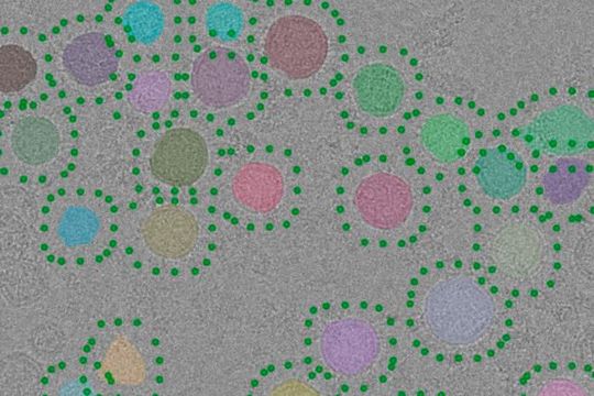
Experimental electron micrograph of purified synaptic vesicles (upper). Synaptic vesicles automatically identified in the image with a new computer program are highlighted with different colours and candidate positions for V-ATPase are indicated with green dots.
(Source: The Hospital for Sick Children)
All brain activity — from memory and emotion to learning and motor control — is made possible through communication across synapses, the connections between neurons. When this communication is unsuccessful, various conditions such as epilepsy can occur. A neuron is a type of cell that specializes in communicating with other cells by sending out chemical signals, called neurotransmitters, into synapses. In the brain, there are 100 trillion synapses between neurons.
The way neurons communicate has been studied for decades, but research published today in Science showcases models derived from hundreds of thousands of high-resolution images that reveal synaptic function with new clarity.
Led by Dr. John Rubinstein, Senior Scientist in the Molecular Medicine program, and Dr. Claire Coupland, first author and postdoctoral fellow in the Rubinstein Lab, the research team hopes that by capturing images of and modeling how chemicals are released from neurons, they may be able to inform new therapeutic targets that help improve care for children with epilepsy and other neurological conditions.
When communicating, neurons release neurotransmitters into a synapse to be delivered to a receiving neuron. These neurotransmitters are released from small packets called synaptic vesicles. Once a message is received, the neurotransmitters must be reabsorbed and repackaged into new synaptic vesicles to clear out the synapse and make room for the next signal. To facilitate this process, an enzyme called the vesicular-type ATPase (V-ATPase) acts as a pump to drive neurotransmitters into synaptic vesicles. V-ATPase also regulates neurotransmitter release from the vesicles.
“In our research, we learned that the way V-ATPase controls the process of neurotransmitter release from synaptic vesicles is by spontaneously falling apart after the vesicles are loaded. We found that when we filled the synaptic vesicles with neurotransmitters, the V-ATPases split into two parts, which then allows neurotransmitter release,” explains Rubinstein.
By using novel biochemical methods and novel imaging methods supported by the Sick Kids Nanoscale Biomedical Imaging Facility, the scientists were able to isolate synaptic vesicles and obtain images of them. From there, they developed new computational approaches to analyze the images to show the V-ATPase in the vesicles at high resolution — something that has not been done before.
“We created 3D models of the V-ATPase based on images we captured using cryogenic electron microscopy (cryo-EM), a method that images samples at -196°C. Our team saw that V-ATPase interacts with several components of the synaptic vesicle, which contains many proteins and lipids that are involved in neurotransmitter release”, says Rubinstein. “Most surprisingly, we learned that the V-ATPase interacts with a protein called synaptophysin. By weight, synaptophysin is the most abundant synaptic vesicle protein. Until now, its function in neurons was not understood. What we found shows that synaptophysin could be helping to recruit V-ATPase to synaptic vesicles when they initially form.”
Now that the researchers have discovered that V-ATPase interacts with synaptophysin in synaptic vesicles, they are working with Dr. Lu-Yang Wang, a Senior Scientist in the Neurosciences & Mental Health program, to understand the role of this interaction in the brain. They also want to understand how the loading of vesicles leads to the V-ATPase falling apart, and how this process controls the release of neurotransmitters from neurons. In the future, this process could be a therapeutic target for many health conditions, including some kinds of epilepsy.
Date: 08.12.2025
Naturally, we always handle your personal data responsibly. Any personal data we receive from you is processed in accordance with applicable data protection legislation. For detailed information please see our privacy policy.
Consent to the use of data for promotional purposes
I hereby consent to Vogel Communications Group GmbH & Co. KG, Max-Planck-Str. 7-9, 97082 Würzburg including any affiliated companies according to §§ 15 et seq. AktG (hereafter: Vogel Communications Group) using my e-mail address to send editorial newsletters. A list of all affiliated companies can be found here
Newsletter content may include all products and services of any companies mentioned above, including for example specialist journals and books, events and fairs as well as event-related products and services, print and digital media offers and services such as additional (editorial) newsletters, raffles, lead campaigns, market research both online and offline, specialist webportals and e-learning offers. In case my personal telephone number has also been collected, it may be used for offers of aforementioned products, for services of the companies mentioned above, and market research purposes.
Additionally, my consent also includes the processing of my email address and telephone number for data matching for marketing purposes with select advertising partners such as LinkedIn, Google, and Meta. For this, Vogel Communications Group may transmit said data in hashed form to the advertising partners who then use said data to determine whether I am also a member of the mentioned advertising partner portals. Vogel Communications Group uses this feature for the purposes of re-targeting (up-selling, cross-selling, and customer loyalty), generating so-called look-alike audiences for acquisition of new customers, and as basis for exclusion for on-going advertising campaigns. Further information can be found in section “data matching for marketing purposes”.
In case I access protected data on Internet portals of Vogel Communications Group including any affiliated companies according to §§ 15 et seq. AktG, I need to provide further data in order to register for the access to such content. In return for this free access to editorial content, my data may be used in accordance with this consent for the purposes stated here. This does not apply to data matching for marketing purposes.
Right of revocation
I understand that I can revoke my consent at will. My revocation does not change the lawfulness of data processing that was conducted based on my consent leading up to my revocation. One option to declare my revocation is to use the contact form found at https://contact.vogel.de. In case I no longer wish to receive certain newsletters, I have subscribed to, I can also click on the unsubscribe link included at the end of a newsletter. Further information regarding my right of revocation and the implementation of it as well as the consequences of my revocation can be found in the data protection declaration, section editorial newsletter.
Original Publication: High-resolution electron cryomicroscopy of V-ATPase in native synaptic vesicles; Science; DOI:10.1126/science.adp5577




:quality(80)/p7i.vogel.de/wcms/a0/22/a0220cebf4618cef31ba3a3c28411770/0133693882v1.jpeg "Researchers have demonstrated a technique that uses powdered eggshells to produce high-quality magnesium alloys, which have a wide variety of automotive, aerospace and biomedical applications. The eggshells are used as a low-cost, environmentally sustainable alternative to conventional calcium materials, which are manufactured from ore using an energy-intensive process. This photo shows an eggshell next to a rod of egg-shell modified magnesium alloy. (Source: Bharat Gwalani, NC State University)")
:quality(80)/p7i.vogel.de/wcms/83/7b/837b7447733daa56e185cb8f61e8d3bf/0133694425v2.jpeg "Researcher Jinlong Wang stands beside a garbage bin fitted with a solar-powered insert on the lid that efficiently removes food waste smells compared to a garbage bin with a typical lid. (Source: Pan Yan)")
:quality(80)/p7i.vogel.de/wcms/47/aa/47aa3f86498782fc62cba3fc50a423fa/0133690841v2.jpeg "Making new biological discoveries with minimal human intervention is an important advance in self-driving laboratories. (Source: NIH Image Gallery / Chalmers University of Technology)")
:quality(80)/p7i.vogel.de/wcms/09/cc/09cc110fbf9e50608616dde34a418e57/0133688419v2.jpeg "HiPSCs cultured on iMatrix Laminin-511 with Stemfit Basic03. (Source: Amsbio)")
:quality(80)/p7i.vogel.de/wcms/3e/3c/3e3cc29b52fedebf1c97dea23b92a760/0133673542v2.jpeg "Fig.1: The path to accurate pH measurements starts with precise calibration. Use fresh buffers and verify mV response for optimal electrode performance. (Source: METTLER TOLEDO)")
:quality(80)/p7i.vogel.de/wcms/2d/92/2d9231a37601aa58b40d4688f609f004/0133687591v2.jpeg "Horiba’s new CE IVDR certified Yumizen H500 CRP benchtop hematology analyzer delivers a simultaneous complete blood count with 5-part extended differential and rapid CRP whole blood measurement. (Source: Horiba )")
:quality(80)/p7i.vogel.de/wcms/e5/e9/e5e97a3c76cbf47308ce21c4a567a812/0133686320v2.jpeg "The picture shows the Kern PDS precision scale in combination with Kern Balanceconnection software. (Source: Kern & Sohn)")
:quality(80)/p7i.vogel.de/wcms/a8/6e/a86e9b1f1561a67038b6d8a9fe629117/0133465534v2.jpeg "Clean, calibrate, and store pH electrodes properly to ensure accurate, reliable measurements every time. (Source: METTLER TOLEDO)")
:quality(80)/p7i.vogel.de/wcms/d6/ef/d6ef487a821a27ff89287c75b9a94dec/0133693832v1.jpeg "Extent of ice loss at Konkordiaplatz on the Great Aletsch Glacier, canton Valais between 2022 and 2026, compared with the average of measurements taken between 1953 and 1983. (Source: M. Huss / A. Linsbauer)")
:quality(80)/p7i.vogel.de/wcms/ef/7c/ef7cd23997787d020fc225c2677625cf/0133621949v2.jpeg "The team recorded over 1.6 million individual flowers and more than 5,000 plant-pollinator interactions across 58 plant species and 405 pollinator species. (Source: University of Bristol)")
:quality(80)/p7i.vogel.de/wcms/6f/ea/6feaf3edd34eb4a9f1df201dc2e6448d/0133548706v2.jpeg "The researchers studied people’s perceptions, beliefs and knowledge about climate change in two coastal regions, one low-lying island and the other a mainland coast. (Source: P Afna)")
:quality(80)/p7i.vogel.de/wcms/8d/5c/8d5c6c7f897f91d6784825eee20b5584/0133321225v1.jpeg "Empa researcher Christoph Hüglin collects samples of particles that are washed out of the air by rain and snow or settle due to gravity. (Source: Empa)")
:quality(80)/p7i.vogel.de/wcms/e1/f4/e1f48a0c4b6d5c00059370affdc13113/0133694409v2.jpeg "Broccoli growing beneath semi-transparent, colored solar panels at the experimental site at Kärrbo Prästgård farm in Sweden. (Source: Silvia Ma Lu)")
:quality(80)/p7i.vogel.de/wcms/95/3e/953e490f1819f6638bc08fe3d4d43e87/0133566684v1.jpeg "The soy protein isolate the researchers fed to the mice. (Source: KyotoU / Tomoko T. Asai)")
:quality(80)/p7i.vogel.de/wcms/ba/38/ba38179dbcd20fbc3887b2709193d0a8/0133566669v2.jpeg "Consumers in a Cornell study showed no consistent bias against the colour of hybrid red wines, with several ranking among the most visually appealing. (Source: free licensed)")
:quality(80)/p7i.vogel.de/wcms/ac/d1/acd1a8888f6e78206f11f2b4eb33926f/0133566582v1.jpeg "A claim on the front of a box of food products is a hint, not a verdict. The nutrition label on the back – added sugars, sodium, saturated fat, all of it together – still tells the fuller story. (Source: free licensed)")
:quality(80)/p7i.vogel.de/wcms/20/cf/20cfcbf3af8ae6e5d837de9a236d146a/0133694093v1.jpeg "Research findings could help researchers develop personalized treatments for liver disease. (Source: University of Missouri)")
:quality(80)/p7i.vogel.de/wcms/fe/8c/fe8c5f140f67c0f66456686c5a7f68c8/0133693408v2.jpeg "The scientists used single-cell RNA sequencing and genetically engineered mouse models to identify a signaling pathway linking cardiac lymphatics, the heart’s outer lining, and key growth genes that together help regulate cardiac growth. (Source: Unsplash)")
:quality(80)/p7i.vogel.de/wcms/47/2d/472d0c14306eb683ebaa90ce7db42a9a/0133693184v2.jpeg "A human ribosome, shown in gold, translating an mRNA molecule (red). Three tRNAs (orange) help assemble the growing protein chain, shown in silver-white. The image is based on cryo-electron microscopy structures of human ribosomes. (Source: © Adrian Bothe)")
:quality(80)/p7i.vogel.de/wcms/c8/66/c866dcdcc681fb5466b43ac994f5b2d9/0133693165v2.jpeg "The key research findings showed that exposure to parental smoking during childhood was linked to changes in heart structure and poorer cognitive performance in middle age. (Source: Unsplash)")
:quality(80)/images.vogel.de/vogelonline/bdb/1710600/1710675/original.jpg "Discover how to set yourself up for success when performing balance-based density determination of solids such as jewelry and precious metals. (Mettler Toledo)")
:quality(80)/images.vogel.de/vogelonline/bdb/1682500/1682579/original.jpg "The new XPR Analytical balance redesigned to meet the needs of laboratory technicians of working fast and lean. (Mettler Toledo)")
:quality(80)/images.vogel.de/vogelonline/bdb/1677000/1677080/original.jpg "Enhance your weighing accuracy, comfort and productivity with the redesigned XPR Analytical balance. (Mettler Toledo)")
:fill(fff,0)/p7i.vogel.de/companies/69/b7/69b7f63f311d9/logo-bimos-tif--2-.jpeg "logo-bimos-tif--2- (https://www.bimos.com/de-de/)")
:quality(80)/p7i.vogel.de/wcms/40/02/40023ce0d6385120abda9c3b51c698a8/0128355756v2.jpeg "Cells actively help to capture and incorporate influenza viruses. Here, a cell is shown, with a virus in the centre of the image. (Source: Emma Hyde/ ETH Zurich)")
:quality(80)/p7i.vogel.de/wcms/92/3b/923ba38a0b5c46d7a51e3f0089eacfcf/0128184036v2.jpeg "New insights reveal why “feeling sick” prompts the brain to promote rest and isolation. (Source: free licensed)")