A team of engineers and neuroscientists has demonstrated for the first time that human brain organoids implanted in mice have established functional connectivity to the animals’ cortex and responded to external sensory stimuli. The implanted organoids reacted to visual stimuli in the same way as surrounding tissues.
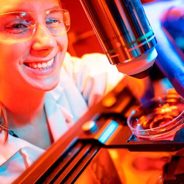
Madison Wilson, a PhD student at UC San Diego, is first author of the study showing that human brain organoids implanted in mice have established functional connectivity to the animals’ cortex and responded to external sensory stimuli.
(Source: David Baillot/ UC San Diego)
Human cortical organoids are derived from human induced pluripotent stem cells, which are usually derived themselves from skin cells. These brain organoids have recently emerged as promising models to study the development of the human brain, as well as a range of neurological conditions.
But until now, no research team had been able to demonstrate that human brain organoids implanted in the mouse cortex were able to share the same functional properties and react to stimuli in the same way. This is because the technologies used to record brain function are limited, and are generally unable to record activity that lasts just a few milliseconds.
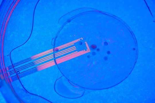
The researchers observed electrical activity in the electrode channels above the organoids showing that the organoids were reacting to the stimulus in the same way as surrounding tissue.
(Source: David Baillot)
A UC San Diego-led team was able to solve this problem by developing experiments that combine microelectrode arrays made from transparent graphene, and two-photon imaging, a microscopy technique that can image living tissue up to one millimeter in thickness.
“No other study has been able to record optically and electrically at the same time,” said Madison Wilson, the paper’s first author and a Ph.D. student in Duygu Kuzum’s research group at UC San Diego. “Our experiments reveal that visual stimuli evoke electrophysiological responses in the organoids, matching the responses from the surrounding cortex.”
The researchers hope that this combination of innovative neural recording technologies to study organoids will serve as a unique platform to comprehensively evaluate organoids as models for brain development and disease, and investigate their use as neural prosthetics to restore function to lost, degenerated or damaged brain regions.
This experimental setup opens up unprecedented opportunities for investigations of human neural network-level dysfunctions underlying developmental brain diseases.
Duygu Kuzum
Kuzum’s lab first developed the transparent graphene electrodes in 2014 and has been advancing the technology since then. The researchers used platinum nanoparticles to lower the impedance of graphene electrodes by 100 times while keeping them transparent. The low-impedance graphene electrodes are able to record and image neuronal activity at both the macroscale and single cell levels.
By placing an array of these electrodes on top of the transplanted organoids, researchers were able to record neural activity electrically from both the implanted organoid and the surrounding host cortex in real time. Using two-photon imaging, they also observed that mouse blood vessels grew into the organoid providing necessary nutrients and oxygen to the implant.
Researchers applied a visual stimulus–an optical white light LED–to the mice with implanted organoids, while the mice were under two-photon microscopy. They observed electrical activity in the electrode channels above the organoids showing that the organoids were reacting to the stimulus in the same way as surrounding tissue. The electrical activity propagated from the area closest to the visual cortex in the implanted organoids area through functional connections. In addition, their low noise transparent graphene electrode technology enabled electrical recording of spiking activity from the organoid and the surrounding mouse cortex. Graphene recordings showed increases in the power of gamma oscillations and phase locking of spikes from organoids to slow oscillations from mouse visual cortex. These findings suggest that the organoids had established synaptic connections with surrounding cortex tissue three weeks after implantation, and received functional input from the mouse brain. Researchers continued these chronic multimodal experiments for eleven weeks and showed functional and morphological integration of implanted human brain organoids with the host mice cortex.
Date: 08.12.2025
Naturally, we always handle your personal data responsibly. Any personal data we receive from you is processed in accordance with applicable data protection legislation. For detailed information please see our privacy policy.
Consent to the use of data for promotional purposes
I hereby consent to Vogel Communications Group GmbH & Co. KG, Max-Planck-Str. 7-9, 97082 Würzburg including any affiliated companies according to §§ 15 et seq. AktG (hereafter: Vogel Communications Group) using my e-mail address to send editorial newsletters. A list of all affiliated companies can be found here
Newsletter content may include all products and services of any companies mentioned above, including for example specialist journals and books, events and fairs as well as event-related products and services, print and digital media offers and services such as additional (editorial) newsletters, raffles, lead campaigns, market research both online and offline, specialist webportals and e-learning offers. In case my personal telephone number has also been collected, it may be used for offers of aforementioned products, for services of the companies mentioned above, and market research purposes.
Additionally, my consent also includes the processing of my email address and telephone number for data matching for marketing purposes with select advertising partners such as LinkedIn, Google, and Meta. For this, Vogel Communications Group may transmit said data in hashed form to the advertising partners who then use said data to determine whether I am also a member of the mentioned advertising partner portals. Vogel Communications Group uses this feature for the purposes of re-targeting (up-selling, cross-selling, and customer loyalty), generating so-called look-alike audiences for acquisition of new customers, and as basis for exclusion for on-going advertising campaigns. Further information can be found in section “data matching for marketing purposes”.
In case I access protected data on Internet portals of Vogel Communications Group including any affiliated companies according to §§ 15 et seq. AktG, I need to provide further data in order to register for the access to such content. In return for this free access to editorial content, my data may be used in accordance with this consent for the purposes stated here. This does not apply to data matching for marketing purposes.
Right of revocation
I understand that I can revoke my consent at will. My revocation does not change the lawfulness of data processing that was conducted based on my consent leading up to my revocation. One option to declare my revocation is to use the contact form found at https://contact.vogel.de. In case I no longer wish to receive certain newsletters, I have subscribed to, I can also click on the unsubscribe link included at the end of a newsletter. Further information regarding my right of revocation and the implementation of it as well as the consequences of my revocation can be found in the data protection declaration, section editorial newsletter.
Next steps include longer experiments involving neurological disease models, as well as incorporating calcium imaging in the experimental set up to visualize spiking activity in organoid neurons. Other methods could also be used to trace axonal projections between organoid and mouse cortex.
“We envision that, further along the road, this combination of stem cells and neurorecording technologies will be used for modeling disease under physiological conditions; examining candidate treatments on patient-specific organoids; and evaluating organoids’ potential to restore specific lost, degenerated or damaged brain regions,” Kuzum said.
References: Multimodal monitoring of human cortical organoids implanted in mice reveal functional connection with visual cortex; Nature Communications; DOI:10.1038/s41467-022-35536-3




:quality(80)/p7i.vogel.de/wcms/62/32/62327aaae1415beeed47db6201e79661/0133375912v2.jpeg "A cotton trailer outfitted with benches and a sunshade offers farmworkers a reprieve from the sun as they take a break from picking cantaloupe melons in Firebaugh, Calif.
(Source: Joe Proudman/ Regents of the University of California)")
:quality(80)/p7i.vogel.de/wcms/e6/c8/e6c8bd554776ee1ad2f1f0723fb243a7/0133369387v1.jpeg "Roman road (Source: Queen Mary University of London)")
:quality(80)/p7i.vogel.de/wcms/51/68/51687ac4cc19f2757c138498b877de57/0133167039v2.jpeg "CMFs are foams that consist of hollow spheres – made of materials such as stainless steel, nickel, or other metals and alloys – embedded in a metallic matrix. The resulting material is both lightweight and remarkably strong at absorbing compressive forces. (Source: Jie Sun/ NC State University)")
:quality(80)/p7i.vogel.de/wcms/cd/c0/cdc09efd1e5b3b8f83224e7729d66c10/0133105276v2.jpeg "From the data presented in this report, the authors show how TF3-MALS-RI is uniquely able to separate large polyacrylate molecules and provide precise molecular weight and Rg values. (Source: Postnova Analytics)")
:quality(80)/p7i.vogel.de/wcms/86/01/86014d3d2a28cefc852f65c8d68317e4/0133104231v2.jpeg "Single-handed tube reading using a Handheld 2. (Source: Azenta Life Sciences)")
:quality(80)/p7i.vogel.de/wcms/0b/0f/0b0f8ed042dfc91f32582e10d54c2c7e/0133087444v2.jpeg "Offering advanced automation to increase your throughput, the Intellixcap provides secure sealing – eliminating the need for manual intervention, while preserving vital sample integrity. (Source: Azenta Life Sciences)")
:quality(80)/p7i.vogel.de/wcms/13/d3/13d35105f74a1c2e22b9c7a270e8e9b3/0132611155v2.jpeg "Circulating cell free DNA. (Source: Amsbio)")
:quality(80)/p7i.vogel.de/wcms/bc/ed/bced6850297a04dbea471a66cb6bb6fe/0132611405v2.jpeg "The latest addition to the range is a peristaltic tubing stop adapter that eliminates the need for fixed stoppers and allows custom cut lengths of peristaltic tubing to be installed on any pump head size. (Source: Biotech Fluidics )")
:quality(80)/p7i.vogel.de/wcms/8d/5c/8d5c6c7f897f91d6784825eee20b5584/0133321225v1.jpeg "Empa researcher Christoph Hüglin collects samples of particles that are washed out of the air by rain and snow or settle due to gravity. (Source: Empa)")
:quality(80)/p7i.vogel.de/wcms/a1/af/a1afbc7c8a067bbe94749ec680b4bafd/0133163762v2.jpeg "The study found in murkier water, fish took longer to move towards the food than those in clear water, but individuals in larger groups responded more quickly overall. (Source: Christos Ioannou)")
:quality(80)/p7i.vogel.de/wcms/32/80/3280aa3d44708962943c96fe2d466744/0133088560v2.jpeg "A bumble bee in flight at the University of Bristol Botanic Garden. (Source: Phil Savoie)")
:quality(80)/p7i.vogel.de/wcms/7b/e5/7be5c735140cbe28f3af0ec3d88274b9/0133045403v2.jpeg "After being buried in the ground for two months and broken down by soil organisms, a new pair of cotton underwear (left) shows clear signs of decomposition (right). (Source: Nicolas Zonvi)")
:quality(80)/p7i.vogel.de/wcms/ee/ad/eead38ab1e3c36f262bd889dfc8c5039/0133409909v2.jpeg "Eating a diet rich in wholegrain food is likely to prevent heart disease by reducing body weight, blood pressure and the levels of cholesterol and glucose in the blood. (Source: Unsplash)")
:quality(80)/p7i.vogel.de/wcms/ef/68/ef68375729607984e5bb0c143afdae70/0133375119v2.jpeg "Printed Rice-Lentil 3D structures
(Source: SUTD)")
:quality(80)/p7i.vogel.de/wcms/bf/d0/bfd087349a85956a7aefad57ac1204c7/0133241961v2.jpeg "The Thermo Scientific Suretect Beverage Spoilage Multiplex PCR Assay provides a fast, broad-range molecular solution for detecting key spoilage microorganisms that threaten beverage quality. (Source: © OlegDoroshin - stock.adobe.com)")
:quality(80)/p7i.vogel.de/wcms/d4/61/d461ce0bd5743897993c6475f69e7ad6/0133067478v2.jpeg "In Harris County, 50 % of households with children reported food insecurity, compared with 33 % of households without children. (Source: Rice University)")
:quality(80)/p7i.vogel.de/wcms/f5/d3/f5d3af7b7479d0573bec5c1fb9f1fd7b/0133409927v2.jpeg "The Match study looked at over 500 cancer survivors with varying types and stages of cancer and found that patients experienced less pain, fatigue, and sleep problems after participating in the interventions. (Source: Unsplash)")
:quality(80)/p7i.vogel.de/wcms/22/49/224964dd6850b62cccdbc4bde2d34693/0133372964v1.jpeg "Brain activity recorded during a video game helped researchers predict whether participants would take or avoid a risk half a second before they acted. (Source: free licensed)")
:quality(80)/p7i.vogel.de/wcms/48/a7/48a761934161543f70fef2403b44d714/0133372328v2.jpeg "The link between pollution and coughing persisted in the data, even when pollution levels were below the WHO’s recommended safe daily exposure limit. (Source: free licensed)")
:quality(80)/images.vogel.de/vogelonline/bdb/1710600/1710675/original.jpg "Discover how to set yourself up for success when performing balance-based density determination of solids such as jewelry and precious metals. (Mettler Toledo)")
:quality(80)/images.vogel.de/vogelonline/bdb/1682500/1682579/original.jpg "The new XPR Analytical balance redesigned to meet the needs of laboratory technicians of working fast and lean. (Mettler Toledo)")
:quality(80)/images.vogel.de/vogelonline/bdb/1677000/1677080/original.jpg "Enhance your weighing accuracy, comfort and productivity with the redesigned XPR Analytical balance. (Mettler Toledo)")
:fill(fff,0)/images.vogel.de/vogelonline/companyimg/107800/107832/65.jpg "logoDueperthal.jpg ()")
:quality(80)/p7i.vogel.de/wcms/83/90/839049b296905df2a07deb4e867ff2cb/0129594701v2.jpeg "The full device next to a quarter for scale. (Source: John A. Rogers/Northwestern University)")
:quality(80)/p7i.vogel.de/wcms/22/49/224964dd6850b62cccdbc4bde2d34693/0133372964v1.jpeg "Brain activity recorded during a video game helped researchers predict whether participants would take or avoid a risk half a second before they acted. (Source: free licensed)")