Alzheimer's is often associated with so-called plaques in the brain. However, these protein deposits on neurons are probably not solely responsible for the disease. A new study by researchers at the Ruhr University Bochum, for example, provides evidence that certain protein residues can also penetrate into the interior of the cells and lead to their death.
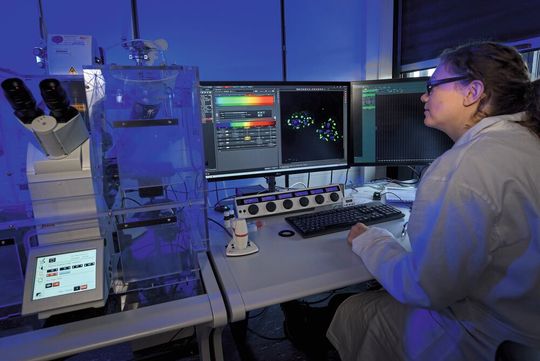
Sophia Meermeyer examines a cell culture sample under the microscope. Various aggregates have formed in the cell nuclei, which are colored blue and yellow, respectively. The yellow aggregates result from the displacement of FE65 and the part of APP that was inside the cell into the nucleus.
(Source: Roberto Schirdewahn)
Bochum/Germany — In addition to plaques that accumulate outside of nerve cells in the brain, Alzheimer’s disease is also characterised by changes inside these cells. Researchers from the Cell Signalling research group at the Chair of Molecular Biochemistry at Ruhr University Bochum (RUB), headed by Dr. Thorsten Müller, have been studying what exactly happens in these cells. They determined that various proteins and protein components accumulate in the cells, which also affect their functions. Moreover, they identified a correlation between the progression of the disease and certain corpuscles in the cell nuclei. They published their report in the journal Acta Neuropathological Communications on 13 April 2021.
Affecting over 50 million people, Alzheimer’s disease is the most common form of dementia and primarily occurs in people over the age of 65. The pathology of the disease in the brain is mainly characterised by two factors:
Beta-amyloid plaques outside the nerve cells: a protein present in the body that is formed by the cleavage of the amyloid precursor protein (APP).
Tau proteins: This protein stabilises tube-like structures (microtubules) inside cells, which are relevant for the transport of nutrients in nerve cells.
APP is embedded in the cell membrane of the nerve cells and protrudes both on the inside and outside. Typically, it is cleaved once near the cell membrane The part inside the nerve cells is unstable and disintegrates. In Alzheimer’s patients, two cleavages take place, resulting in the formation of three parts. The new findings now indicate that the part inside the nerve cells is more stable in Alzheimer’s patients. It consists of only approximately 50 amino acids and can, under certain conditions, migrate into the cell nucleus together with other proteins such as FE65 and TIP60. This protein complex, also known as nuclear aggregates, has the capacity to manipulate the cell’s gene expression. “This suggests that the aggregates have a function in this region,” points out lead author David Marks from the Cell Signaling group at RUB. Closer analysis confirmed this evidence, as a protein was found within the nuclear aggregates that is involved in the modification of DNA.
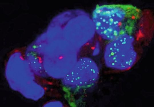
The green dots are protein aggregates in the cell nuclei. In cells that appear solid blue, no aggregates have formed (yet).
(Source: Thorsten Müller)
Two More Protein Candidates in Cells
“To understand this mechanism even better, we looked for other proteins that might be part of these aggregates and identified two more candidates involved in the nuclear aggregates, the so-called tumour suppressor proteins P53 and PML,” explains David Marks. The study team showed in experiments on living cells that nuclear aggregates formed from the proteins APP-CT50, FE65, TIP60 and PML fuse with each other over time and produce even larger nuclear aggregates.
In addition, they examined brain samples from older deceased persons as well as self-generated neuronal tissue from induced pluripotent stem cells. “These mini-brains, so-called cerebral organoids, reflect the embryonic and developmental phase of a brain fairly accurately,” explains Thorsten Müller. The research team found nearly no nuclear aggregates in the comparatively young cerebral organoids, whereas they were present in brain samples from older patients. “This leads us to conclude that the process depends on age,” says Müller.
Furthermore, the researchers showed on these brain samples that APP-CT50 and FE65 can make up part of the so-called PML bodies, which occur naturally in large numbers of cell nuclei. Upon further analysis of hippocampal brain slices from Alzheimer’s patients, the authors identified a significant correlation between a reduced number of PML bodies in the nucleus in regions with a high beta-amyloid plaque load. In particular, such regions mean that there is higher expression and processing of APP. This may suggest that the transfer of APP-CT50 and FE65 into the nucleus is part of AD pathology and affects the fusion of PML bodies there.
Date: 08.12.2025
Naturally, we always handle your personal data responsibly. Any personal data we receive from you is processed in accordance with applicable data protection legislation. For detailed information please see our privacy policy.
Consent to the use of data for promotional purposes
I hereby consent to Vogel Communications Group GmbH & Co. KG, Max-Planck-Str. 7-9, 97082 Würzburg including any affiliated companies according to §§ 15 et seq. AktG (hereafter: Vogel Communications Group) using my e-mail address to send editorial newsletters. A list of all affiliated companies can be found here
Newsletter content may include all products and services of any companies mentioned above, including for example specialist journals and books, events and fairs as well as event-related products and services, print and digital media offers and services such as additional (editorial) newsletters, raffles, lead campaigns, market research both online and offline, specialist webportals and e-learning offers. In case my personal telephone number has also been collected, it may be used for offers of aforementioned products, for services of the companies mentioned above, and market research purposes.
Additionally, my consent also includes the processing of my email address and telephone number for data matching for marketing purposes with select advertising partners such as LinkedIn, Google, and Meta. For this, Vogel Communications Group may transmit said data in hashed form to the advertising partners who then use said data to determine whether I am also a member of the mentioned advertising partner portals. Vogel Communications Group uses this feature for the purposes of re-targeting (up-selling, cross-selling, and customer loyalty), generating so-called look-alike audiences for acquisition of new customers, and as basis for exclusion for on-going advertising campaigns. Further information can be found in section “data matching for marketing purposes”.
In case I access protected data on Internet portals of Vogel Communications Group including any affiliated companies according to §§ 15 et seq. AktG, I need to provide further data in order to register for the access to such content. In return for this free access to editorial content, my data may be used in accordance with this consent for the purposes stated here. This does not apply to data matching for marketing purposes.
Right of revocation
I understand that I can revoke my consent at will. My revocation does not change the lawfulness of data processing that was conducted based on my consent leading up to my revocation. One option to declare my revocation is to use the contact form found at https://contact.vogel.de. In case I no longer wish to receive certain newsletters, I have subscribed to, I can also click on the unsubscribe link included at the end of a newsletter. Further information regarding my right of revocation and the implementation of it as well as the consequences of my revocation can be found in the data protection declaration, section editorial newsletter.




:quality(80)/p7i.vogel.de/wcms/62/32/62327aaae1415beeed47db6201e79661/0133375912v2.jpeg "A cotton trailer outfitted with benches and a sunshade offers farmworkers a reprieve from the sun as they take a break from picking cantaloupe melons in Firebaugh, Calif.
(Source: Joe Proudman/ Regents of the University of California)")
:quality(80)/p7i.vogel.de/wcms/e6/c8/e6c8bd554776ee1ad2f1f0723fb243a7/0133369387v1.jpeg "Roman road (Source: Queen Mary University of London)")
:quality(80)/p7i.vogel.de/wcms/51/68/51687ac4cc19f2757c138498b877de57/0133167039v2.jpeg "CMFs are foams that consist of hollow spheres – made of materials such as stainless steel, nickel, or other metals and alloys – embedded in a metallic matrix. The resulting material is both lightweight and remarkably strong at absorbing compressive forces. (Source: Jie Sun/ NC State University)")
:quality(80)/p7i.vogel.de/wcms/cd/c0/cdc09efd1e5b3b8f83224e7729d66c10/0133105276v2.jpeg "From the data presented in this report, the authors show how TF3-MALS-RI is uniquely able to separate large polyacrylate molecules and provide precise molecular weight and Rg values. (Source: Postnova Analytics)")
:quality(80)/p7i.vogel.de/wcms/86/01/86014d3d2a28cefc852f65c8d68317e4/0133104231v2.jpeg "Single-handed tube reading using a Handheld 2. (Source: Azenta Life Sciences)")
:quality(80)/p7i.vogel.de/wcms/0b/0f/0b0f8ed042dfc91f32582e10d54c2c7e/0133087444v2.jpeg "Offering advanced automation to increase your throughput, the Intellixcap provides secure sealing – eliminating the need for manual intervention, while preserving vital sample integrity. (Source: Azenta Life Sciences)")
:quality(80)/p7i.vogel.de/wcms/13/d3/13d35105f74a1c2e22b9c7a270e8e9b3/0132611155v2.jpeg "Circulating cell free DNA. (Source: Amsbio)")
:quality(80)/p7i.vogel.de/wcms/bc/ed/bced6850297a04dbea471a66cb6bb6fe/0132611405v2.jpeg "The latest addition to the range is a peristaltic tubing stop adapter that eliminates the need for fixed stoppers and allows custom cut lengths of peristaltic tubing to be installed on any pump head size. (Source: Biotech Fluidics )")
:quality(80)/p7i.vogel.de/wcms/8d/5c/8d5c6c7f897f91d6784825eee20b5584/0133321225v1.jpeg "Empa researcher Christoph Hüglin collects samples of particles that are washed out of the air by rain and snow or settle due to gravity. (Source: Empa)")
:quality(80)/p7i.vogel.de/wcms/a1/af/a1afbc7c8a067bbe94749ec680b4bafd/0133163762v2.jpeg "The study found in murkier water, fish took longer to move towards the food than those in clear water, but individuals in larger groups responded more quickly overall. (Source: Christos Ioannou)")
:quality(80)/p7i.vogel.de/wcms/32/80/3280aa3d44708962943c96fe2d466744/0133088560v2.jpeg "A bumble bee in flight at the University of Bristol Botanic Garden. (Source: Phil Savoie)")
:quality(80)/p7i.vogel.de/wcms/7b/e5/7be5c735140cbe28f3af0ec3d88274b9/0133045403v2.jpeg "After being buried in the ground for two months and broken down by soil organisms, a new pair of cotton underwear (left) shows clear signs of decomposition (right). (Source: Nicolas Zonvi)")
:quality(80)/p7i.vogel.de/wcms/ee/ad/eead38ab1e3c36f262bd889dfc8c5039/0133409909v2.jpeg "Eating a diet rich in wholegrain food is likely to prevent heart disease by reducing body weight, blood pressure and the levels of cholesterol and glucose in the blood. (Source: Unsplash)")
:quality(80)/p7i.vogel.de/wcms/ef/68/ef68375729607984e5bb0c143afdae70/0133375119v2.jpeg "Printed Rice-Lentil 3D structures
(Source: SUTD)")
:quality(80)/p7i.vogel.de/wcms/bf/d0/bfd087349a85956a7aefad57ac1204c7/0133241961v2.jpeg "The Thermo Scientific Suretect Beverage Spoilage Multiplex PCR Assay provides a fast, broad-range molecular solution for detecting key spoilage microorganisms that threaten beverage quality. (Source: © OlegDoroshin - stock.adobe.com)")
:quality(80)/p7i.vogel.de/wcms/d4/61/d461ce0bd5743897993c6475f69e7ad6/0133067478v2.jpeg "In Harris County, 50 % of households with children reported food insecurity, compared with 33 % of households without children. (Source: Rice University)")
:quality(80)/p7i.vogel.de/wcms/f5/d3/f5d3af7b7479d0573bec5c1fb9f1fd7b/0133409927v2.jpeg "The Match study looked at over 500 cancer survivors with varying types and stages of cancer and found that patients experienced less pain, fatigue, and sleep problems after participating in the interventions. (Source: Unsplash)")
:quality(80)/p7i.vogel.de/wcms/22/49/224964dd6850b62cccdbc4bde2d34693/0133372964v1.jpeg "Brain activity recorded during a video game helped researchers predict whether participants would take or avoid a risk half a second before they acted. (Source: free licensed)")
:quality(80)/p7i.vogel.de/wcms/48/a7/48a761934161543f70fef2403b44d714/0133372328v2.jpeg "The link between pollution and coughing persisted in the data, even when pollution levels were below the WHO’s recommended safe daily exposure limit. (Source: free licensed)")
:quality(80)/images.vogel.de/vogelonline/bdb/1710600/1710675/original.jpg "Discover how to set yourself up for success when performing balance-based density determination of solids such as jewelry and precious metals. (Mettler Toledo)")
:quality(80)/images.vogel.de/vogelonline/bdb/1682500/1682579/original.jpg "The new XPR Analytical balance redesigned to meet the needs of laboratory technicians of working fast and lean. (Mettler Toledo)")
:quality(80)/images.vogel.de/vogelonline/bdb/1677000/1677080/original.jpg "Enhance your weighing accuracy, comfort and productivity with the redesigned XPR Analytical balance. (Mettler Toledo)")
:fill(fff,0)/images.vogel.de/vogelonline/companyimg/107800/107832/65.jpg "logoDueperthal.jpg ()")
:fill(fff,0)/p7i.vogel.de/companies/69/b7/69b7f63f311d9/logo-bimos-tif--2-.jpeg "logo-bimos-tif--2- (https://www.bimos.com/de-de/)")
:quality(80)/images.vogel.de/vogelonline/bdb/1805400/1805472/original.jpg "n this artist’s rendering, amyloid plaques are interspersed among neurons. These aggregates of misfolded proteins disrupt and kill brain cells, and are a hallmark of Alzheimer’s disease. (National Institute of Aging)")
:quality(80)/p7i.vogel.de/wcms/1a/ee/1aee2613e1a69c73ab568ee61d354ded/0129347044v1.jpeg "The Tonks lab studied PTP1B inhibition in a mouse model of Alzheimer’s disease. When PTP1B was deleted, as shown in the bottom row, the brain’s immune cells (green) were better at engulfing harmful amyloid-β plaques (grey), as shown in the left column. (Source: Nicholas Tonks/ CSHL)")
:quality(80)/p7i.vogel.de/wcms/15/e0/15e08e2ebb02e3d0bd13bba1c0ff44e4/0131588088v2.jpeg "In the new study, the scientists examined young and old male and female mice, with or without loss of brain estrogen, which allowed them to pinpoint the effects specifically relevant to older females. (Source: 愚木混株 Yumu on Unsplash)")