

:quality(80)/p7i.vogel.de/wcms/ac/24/ac2495bc671ef2a8bf95bdb0de7f172a/0131908887v1.jpeg "An artistic rendering of a boron nitride nanotube developed by UIC researchers and their collaborators in a new study. (Source: Rendering by Ella Maru Studio, Inc. Courtesy of Sangil Kim)")
:quality(80)/p7i.vogel.de/wcms/dd/be/ddbe621ac35ec49d9aee43615173f703/0131898392v2.jpeg "The acquisition is consistent with GSK’s strategy of acquiring assets that have validated targets and meaningfully address efficacy and/or tolerability limitations of existing standard-of-care therapies. (Source: Pixabay)")
:quality(80)/p7i.vogel.de/wcms/e2/ae/e2aea4192de2e22dd33d19afee25f312/0131779278v1.jpeg "The artificial photosynthesis system produces formic acid from carbon dioxide and water. The right image shows the group’s unique electrolyzer.
(Source: Osaka Metropolitan University)")
:quality(80)/p7i.vogel.de/wcms/49/03/49033fd7e1cb79cd90776e8d0583b441/0131773380v1.jpeg "Taken from above, this annotated image shows a schoolground in Spain and the motions of teenage participants as indicated by the dots and lines. (Source: Echeverría-Huarte et al.)")
:quality(80)/p7i.vogel.de/wcms/f5/8d/f58d7181bd0660dea9e33b89f15947eb/0131589395v2.jpeg "Equipping all your HPLC systems running standard separation protocols with an automated non-invasive flow monitoring system, such as Flowchrom, is the only true solution. (Source: Testa Analytical)")
:quality(80)/p7i.vogel.de/wcms/63/44/63443a618001fbdc12e18de386a7b548/0131588936v2.jpeg "These compact, chemically inert degassers use trusted Degasi degasser parts, combined in a manner optimized to your specific system requirements. (Source: Biotech Fluidics)")
:quality(80)/p7i.vogel.de/wcms/47/13/4713969ee1fed4c9d6cd932df7c82482/0131588920v2.jpeg "My Genius Pro installed at a leading European transplant center supporting routine infectious disease testing. (Source: Elitech Group)")
:quality(80)/p7i.vogel.de/wcms/45/d9/45d9cf5be3c10b389fd712f319fbfce1/0131588911v2.jpeg "The Nexis GC-2060 supports both routine and ad-hoc analyses through a range of workflow support functions tailored to operational needs – from automating analytical tasks to on-demand operation that analyses exactly what is needed, when it is needed. (Source: Shimadzu)")
:quality(80)/p7i.vogel.de/wcms/b6/6a/b66a4a25066ff8e426cd9f574b72c7b9/0131905275v1.jpeg "PLA plastic (left piece) usually takes months to break down in industrial composting facilities, but incorporating a sprinkling of an organic additive dramatically speeds the process for a modified PLA plastic (right piece) to less than three weeks.
(Source: Jinsol Yook)")
:quality(80)/p7i.vogel.de/wcms/9f/d8/9fd8ffb67c1132b4f48dc60e4e194226/0131784166v2.jpeg "Still life featuring protein beads loaded with potassium hydroxide. The porous act as a sponge for CO2. (Source: Mezzenga Lab / ETH Zurich)")
:quality(80)/p7i.vogel.de/wcms/3e/47/3e472068bf13aadf4786e947ae148759/0131779258v2.jpeg "Salts, organic matter and other chemical constituents in natural fresh water and seawater suppress sunlight's ability to degrade plastic. Because sunlight cannot effectively initiate the degradation process, microbes cannot finish the job. That means nature’s cleanup process slows down, allowing plastics to accumulate and persist in waterways around the world. (Source: Ludmilla Aristilde/ Northwestern University)")
:quality(80)/p7i.vogel.de/wcms/bc/bc/bcbcf8be4b6c996f6ff231cb65937953/0131673749v1.jpeg "Sewage overflows occur when untreated sewage enters homes or the environment through broken or clogged pipes, or when the sewage system is overwhelmed. (Source: free licensed)")
:quality(80)/p7i.vogel.de/wcms/ac/33/ac339ce19b25f78e11a8dc8ef137869b/0131904977v2.jpeg "An “electronic nose” created by UC Berkeley researchers can detect the gases emitted by spoiled food and food allergens better than human noses. (Source: Brandon Sánchez-Mejia/ UC Berkeley)")
:quality(80)/p7i.vogel.de/wcms/b2/f9/b2f99d68013e2a6ab65e6c86254ec8a9/0131802730v2.jpeg "The UK currently eats 31 % less seafood than is recommended by government guidelines. (Source: University of Bristol)")
:quality(80)/p7i.vogel.de/wcms/1e/ed/1eed2310aac6373fe3d57c8f9d2809d6/0131673857v2.jpeg "“Addressing structural and policy-related barriers to accessing fresh and minimally processed foods remains critical for promoting dietary changes that improve the health and life span for all Americans,” said Dariush Mozaffarian, cardiologist and director of the Food is Medicine Institute at the Friedman School of Nutrition Science and Policy at Tufts University.
(Source: Imani Khayaam for Tufts University)")
:quality(80)/p7i.vogel.de/wcms/48/97/4897129d96e4ab9287933406b153ab51/0131163956v2.jpeg "Eating one egg per day for at least five days a week reduces risk of Alzheimer’s by up to 27%, researchers found. (Source: free licensed)")
:quality(80)/p7i.vogel.de/wcms/17/20/1720579fad3f2e89bc1e190e42ee7c8f/0131911326v2.jpeg "Wild cotton, on left, has short, brown, and coarse fibers, while modern domesticated cotton has white, fine and abundant fibers. A new study led by Iowa State University scientists identified the northwestern Yucatan Peninsula in Mexico as the original source of domesticated cotton. (Source: Corrinne Grover/ Iowa State Universit)")
:quality(80)/p7i.vogel.de/wcms/e0/47/e0471aba187b684e713df3c4d63461f8/0131909497v1.jpeg "Banded mongooses (Mungos mungo) can cooperate with common warthogs (Phacochoerus africanus) by cleaning them, removing ticks and other parasites, while the warthogs provide access to food and safety from predators through their vigilance and presence. Example footage from Queen Elizabeth National Park, Uganda.
(Source: Leela Channer)")
:quality(80)/p7i.vogel.de/wcms/db/28/db28c59875bbfbeae78aac866c08ffa3/0131901880v2.jpeg "Scientists at Northwestern University and Shirley Ryan Abilitylab have developed a first-of-its-kind rehabilitation system that virtually connects therapists and patients through robotic exoskeletons. (Source: Shirley Ryan Abilitylab)")
:quality(80)/p7i.vogel.de/wcms/bb/51/bb51f90e36d72be4dd360c026642b09c/0131843656v2.jpeg "As patients live longer, they are at an increased risk of developing valvular heart disease after successful cancer therapy. (Source: Pixabay)")
:quality(80)/images.vogel.de/vogelonline/bdb/1710600/1710675/original.jpg "Discover how to set yourself up for success when performing balance-based density determination of solids such as jewelry and precious metals. (Mettler Toledo)")
:quality(80)/images.vogel.de/vogelonline/bdb/1682500/1682579/original.jpg "The new XPR Analytical balance redesigned to meet the needs of laboratory technicians of working fast and lean. (Mettler Toledo)")
:quality(80)/images.vogel.de/vogelonline/bdb/1677000/1677080/original.jpg "Enhance your weighing accuracy, comfort and productivity with the redesigned XPR Analytical balance. (Mettler Toledo)")
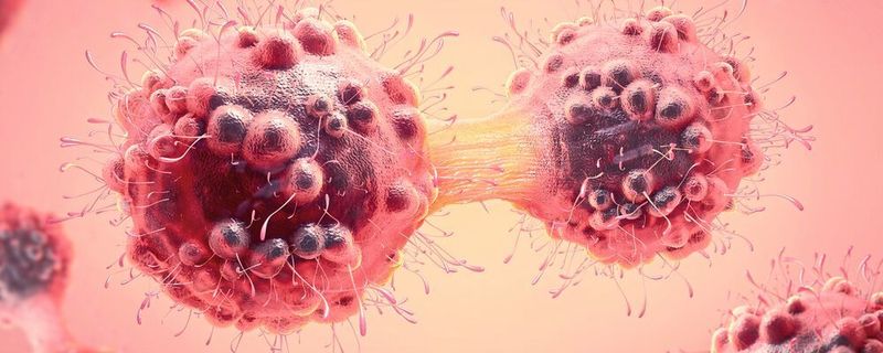
3D ex vivo Microfluidics Platform Beyond Limitations: New Microfluidics Platform for Cancer Drug Screening and Profiling
High-attrition rates during clinical development of new cancer therapies still persist. A new 3D ex vivo microfluidics platform shall now enhance cancer drug screening and early development process by creating cancer models which predict clinical outcomes with significantly increased reliability.
Related Vendor
:fill(fff,0)/images.vogel.de/vogelonline/companyimg/131900/131910/65.jpg "Xylem_tag_rgb.jpg ()")
Cancer is one of the leading causes of mortality. Specific cancer types, such as glioma and pancreatic, remain intractable to new therapies and advanced cancers represent critical areas of unmet therapeutic need. The challenges in developing successful cancer therapeutics is not just limited to the discovery of new drug therapies, but also to the availability of robust preclinical ex vivo models. Recent technological advances in high-throughput and content phenotypic screening, as well as 3D multicellular assay methods, have opened doors for reshaping several key processes during drug discovery and development.
Challenges in cancer therapeutics
Despite increased research investments and ongoing efforts toward finding novel modalities for the treatment of the most challenging cancers, translation into patient benefit has been slow. Progress from preclinical drug discovery to positive clinical outcomes is limited by the fact that translational cancer research from early drug discovery to late stage drug development and assessment in clinical trials is a long process. Moreover, traditional drug discovery and discovery medicine activities have relied upon rudimentary 2D cell monolayer models , 3D clonogenic assay platforms and small animal models, mostly utilizing established cancer cell lines.
![(extracted from Xin X, et al (2016), Cell Stem Cell [19])](https://cdn1.vogel.de/YW-JiwA5XXrtp48KrOy5Q08S-9Y=/300x300/smart/filters:format(jpg):quality(80)/images.vogel.de/vogelonline/bdb/1401900/1401964/original.jpg "(extracted from Xin X, et al (2016), Cell Stem Cell [19])")
:quality(80)/images.vogel.de/vogelonline/bdb/1401900/1401965/original.jpg "(Amsbio)")
It is now apparent that such preclinical models do not accurately recapitulate the complex pathophysiology of cancers found in patients and poorly predict clinical response. Therefore, development of more predictive, patient-specific models of human cancer are pivotal in the quest for profiling of novel anticancer drugs, either as single entities or as drug combinations. Oncologists have recognized the biological similarities of cells grown ex vivo in 3D culture systems with avascular tumors in vivo several decades ago [1, 2). The better the in-vitro models reflect the function and structure of their in-vivo counter parts, the more predictive the cell-based assays becomes. In this regard, several advances have been made in developing robust ex vivo 3D cell culture platforms using microtiter plate formats, 3D perfusion systems and 3D microfluidics formats with special focus on multicellular tumor spheroids (MCTS) systems. The emergence of these technologies have made it possible to conduct drug screening and early development programs in cost effective and efficient ways using clinically relevant cell types derived from multiple cancers.
Currently, the use of ex vivo 3D assay platforms is being pursued by researchers as a tool for drug activity response and monitoring potential changes at subcellular levels to identify downstream targets post drug treatment. Additionally, these platforms are extensively being used as a drug development tool as it has been shown to closely simulate the tumour microenvironment within the in vitro milieu [3-6]. It has previously been reported that the drug response of cancer cells is not just determined by the inherent characteristics of the epithelial tumor cells, but, are also controlled by signals derived from other cells (stromal/fibroblasts/distinct immune cell compartments) within the tumor microenvironment [7-10].
Harris AL (2002) [11] and Mellor & Callaghan (2008) [12] have reported that the abnormal vascularization of solid tumors leads to the generation of tumor microenvironments that are chronically starved of oxygen and nutrients. As a result, cells residing in such environment demonstrate altered phenotypic characteristics when compared to the cells located in more vascularized regions around the outer core [13-15]. Therefore, there has been immense drive towards developing methods which can exploit the altered phenotype of tumors.
One of the earliest 3D-related effects, which were correlated to in-vivo observations, included multicellular drug resistance (MDR). Cancer cells grown as 3D spheroids generally display reduced drug sensitivity compared to 2D monolayer cultures [16]. These observations indicate that the use of 3D spherical microtissues may enable improved discrimination of the most active anti-cancer drugs with improved therapeutic index.
(ID:45276983)
:quality(80)/p7i.vogel.de/wcms/a1/fa/a1fa8b58dec1480da3b20c7b9ffabb2d/0131301715v2.jpeg "By tagging nanoparticles with unique DNA “barcodes”, the NUS research team was able to track and compare dozens of designs simultaneously in living tumor models, rapidly identifying those most effective at reaching mitochondria (energy centres inside cancer cells). (Source: NUS)")
:quality(80)/p7i.vogel.de/wcms/6e/b4/6eb490e9ca40cb42034b3052a874e46e/0129778274v2.jpeg "The researchers tested a specific class of these PLPs, called Hydracs (Hybrid Degrading Copolymers), on two particularly problematic proteins: MYC and KRAS. (Source: Nathan Gianneschi/Northwestern University)")